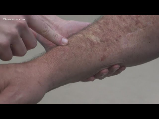
Melanoma Cancer Pictures refer to images that visually depict melanoma, a serious form of skin cancer. One common example is a dark, irregular mole with jagged edges and multiple colors.
These pictures play a crucial role in raising awareness and facilitating early detection of melanoma. They help individuals identify potential warning signs and seek medical attention promptly. Historically, the advent of digital photography and the internet have significantly increased the accessibility of these images, making them widely available for educational purposes.
In this article, we will explore various types of melanoma cancer pictures, their diagnostic value, and the importance of using this visual information to improve melanoma patient outcomes.
Melanoma Cancer Pictures
Melanoma cancer pictures play a vital role in early detection, diagnosis, and public awareness. They provide visual representations of melanoma, enabling individuals to recognize the warning signs and seek medical attention promptly.
- Educational Tool
- Diagnostic Aid
- Awareness Campaign
- Clinical Documentation
- Research Resource
- Telemedicine Consultation
- Patient Education
- Public Health Campaign
- Social Media Advocacy
- Melanoma Screening
These various aspects highlight the significance of melanoma cancer pictures in raising awareness, facilitating diagnosis, supporting research, and ultimately improving patient outcomes. They serve as a valuable educational tool for healthcare professionals and the general public, empowering individuals to take an active role in their skin health and potentially saving lives.
Educational Tool
Melanoma cancer pictures serve as a powerful educational tool, enabling individuals to recognize and identify the warning signs of melanoma, a serious form of skin cancer. These visual representations provide a comprehensive understanding of melanoma's characteristics, aiding in early detection and facilitating timely medical intervention.
Educational institutions and healthcare organizations utilize melanoma cancer pictures extensively in teaching materials, awareness campaigns, and patient education programs. The vivid imagery helps students, healthcare professionals, and the general public grasp the variations in melanoma's appearance, including its diverse colors, shapes, and sizes. By raising awareness and providing visual references, melanoma cancer pictures empower individuals to perform self-examinations and seek medical attention if suspicious lesions are detected.
The practical applications of melanoma cancer pictures extend beyond medical settings. Public health campaigns leverage these images to educate communities about the importance of sun protection and regular skin checks. Social media platforms and online resources disseminate melanoma cancer pictures, reaching a wide audience and fostering a culture of skin cancer awareness.
In summary, melanoma cancer pictures are indispensable educational tools, playing a pivotal role in early detection, public awareness, and patient education. Their ability to visually convey complex medical information empowers individuals to take an active role in their skin health, ultimately leading to improved melanoma outcomes.
Diagnostic Aid
Melanoma cancer pictures serve as a valuable diagnostic aid, assisting healthcare professionals in accurately identifying and assessing melanoma lesions. These visual representations provide critical information that complements physical examinations and other diagnostic techniques.
- Visual Assessment: Melanoma cancer pictures enable healthcare providers to visualize the size, shape, color, and distribution of melanoma lesions. This visual information helps in differentiating melanoma from benign lesions and assessing the severity and stage of the disease.
- Telemedicine Consultations: In remote areas or for patients with limited mobility, melanoma cancer pictures facilitate telemedicine consultations. Healthcare providers can remotely evaluate lesions, providing timely advice and guidance, ensuring accessible and convenient care.
- Dermoscopy Analysis: Specialized melanoma cancer pictures using dermoscopy techniques provide magnified views of skin lesions. This detailed visualization aids in identifying subtle patterns and structures, enhancing the accuracy of melanoma diagnosis.
- AI-Assisted Diagnosis: Artificial intelligence algorithms trained on melanoma cancer pictures can assist healthcare providers in evaluating lesions and making diagnostic suggestions. This technology offers a second opinion, reducing diagnostic errors and improving patient outcomes.
In summary, melanoma cancer pictures are indispensable diagnostic aids, empowering healthcare professionals with visual information to accurately identify and assess melanoma lesions. Their role in telemedicine, dermoscopy analysis, and AI-assisted diagnosis further enhances their diagnostic value, leading to improved patient care and management.
Awareness Campaign
In the context of melanoma cancer, awareness campaigns play a pivotal role in disseminating critical information to the public, fostering early detection, and reducing the burden of the disease. Melanoma cancer pictures serve as powerful tools within these campaigns, visually conveying the warning signs and characteristics of melanoma.
- Public Education: Campaigns utilize melanoma cancer pictures to educate the public about the importance of skin protection, regular skin self-examinations, and prompt medical attention for suspicious lesions.
- Community Outreach: Melanoma cancer pictures are instrumental in community outreach programs, bringing awareness to high-risk populations and underserved communities.
- Social Media Advocacy: Social media platforms provide a vast reach for melanoma cancer picture-based campaigns, spreading awareness and promoting skin cancer prevention.
- Targeted Campaigns: Campaigns tailored to specific demographics or geographic regions use melanoma cancer pictures to address local needs and improve early detection rates.
These facets of awareness campaigns, powered by melanoma cancer pictures, collectively contribute to increased public knowledge, improved self-screening practices, and timely medical intervention. By raising awareness and fostering a culture of skin cancer prevention, these campaigns ultimately lead to better melanoma outcomes and reduced mortality rates.
Clinical Documentation
In the context of melanoma cancer, clinical documentation plays a vital role in ensuring accurate and comprehensive patient records. Melanoma cancer pictures serve as a crucial component of this documentation, providing visual evidence and aiding in diagnosis, treatment planning, and follow-up care.
- Patient Records: Melanoma cancer pictures are incorporated into patient medical records, providing a visual representation of the patient's skin lesions over time. This documentation facilitates tracking disease progression, treatment response, and recurrence.
- Diagnostic Tool: Clinical documentation of melanoma cancer pictures assists in the diagnostic process. By capturing the appearance, size, and location of lesions, these pictures aid in differentiating melanoma from other skin conditions, such as benign moles.
- Treatment Planning: Melanoma cancer pictures provide valuable information for treatment planning. The visual documentation helps determine the appropriate treatment approach, including surgical excision, radiation therapy, or systemic therapy.
- Treatment Monitoring: Regular clinical documentation of melanoma cancer pictures enables healthcare providers to monitor treatment response and disease progression. By comparing images over time, they can assess the effectiveness of treatment and make necessary adjustments.
In summary, clinical documentation of melanoma cancer pictures serves as an essential tool for accurate patient records, aiding in diagnosis, treatment planning, and monitoring. This comprehensive documentation contributes to optimal patient care and improves overall melanoma outcomes.
Research Resource
Melanoma cancer pictures serve as a valuable research resource, contributing to advancements in melanoma diagnosis, treatment, and prevention. These images provide a wealth of data for scientific investigation, enabling researchers to study melanoma's characteristics, progression, and response to therapies.
- Disease Classification: Melanoma cancer pictures aid in classifying melanoma subtypes, identifying unique patterns and characteristics that help guide treatment decisions and prognosis.
- Treatment Evaluation: Researchers use melanoma cancer pictures to assess the effectiveness of new and existing treatments. By comparing images before and after treatment, they can evaluate tumor response and identify potential side effects.
- Biomarker Discovery: Melanoma cancer pictures facilitate the discovery of biomarkers, which are indicators of disease presence or progression. Researchers analyze these images to identify specific patterns or features associated with melanoma, leading to the development of new diagnostic and prognostic tools.
- Computational Analysis: Advanced computational techniques, such as machine learning and artificial intelligence, are applied to melanoma cancer pictures to develop algorithms for automated detection, classification, and prediction of melanoma.
The incorporation of melanoma cancer pictures into research studies has significantly contributed to our understanding of melanoma and its management. These images empower researchers to conduct in-depth analyses, leading to advancements in melanoma care and improved patient outcomes.
Telemedicine Consultation
Telemedicine consultation has revolutionized the delivery of healthcare services, including the diagnosis and management of melanoma cancer. Melanoma cancer pictures play a pivotal role in telemedicine consultations, enabling remote evaluation and decision-making.
- Remote Assessment: Patients can send high-quality melanoma cancer pictures to healthcare providers for remote assessment. This eliminates geographical barriers and reduces the need for in-person visits.
- Expert Opinions: Telemedicine consultations allow patients to access opinions from specialized melanoma experts located in different regions or countries. This ensures access to the highest level of expertise, regardless of location.
- Real-Time Collaboration: Telemedicine platforms enable real-time collaboration between healthcare providers and patients. This facilitates interactive discussions, image sharing, and prompt decision-making.
- Improved Accessibility: Telemedicine consultations increase accessibility to melanoma care, especially for patients in underserved areas or with limited mobility. It provides a convenient and efficient way to receive expert medical advice.
The integration of melanoma cancer pictures into telemedicine consultations enhances the accuracy and efficiency of remote diagnosis, expands access to specialized expertise, and improves the overall patient experience. This innovative approach is transforming melanoma care and leading to improved outcomes.
Patient Education
Patient education plays a vital role in melanoma cancer care, empowering individuals with the knowledge and skills to actively participate in their treatment and recovery. Melanoma cancer pictures serve as a powerful tool in patient education, providing visual representations that enhance understanding and promote informed decision-making.
Melanoma cancer pictures enable patients to visualize the characteristics and variations of melanoma, helping them recognize potential warning signs and understand the importance of early detection. Through educational materials, healthcare providers can use these pictures to illustrate the different types of melanoma, their common locations, and the need for regular skin self-examinations.
Real-life examples of patient education using melanoma cancer pictures include interactive online modules, patient support group presentations, and printed educational materials distributed in healthcare settings. These resources leverage high-quality images to demonstrate the diverse clinical presentations of melanoma, emphasizing the importance of prompt medical evaluation for suspicious lesions.
The practical applications of this understanding extend beyond the patient-provider interaction. Public awareness campaigns that incorporate melanoma cancer pictures educate the general population about the risks and signs of melanoma, promoting early detection and reducing the burden of the disease. Social media platforms and online forums also provide opportunities for patients and their families to share experiences, support one another, and learn from the collective knowledge of the community.
Public Health Campaign
Public health campaigns play a crucial role in raising awareness about melanoma cancer and promoting early detection, which is vital for improving patient outcomes. These campaigns often utilize melanoma cancer pictures to convey the warning signs and characteristics of the disease, making them an essential component of public health efforts to combat melanoma.
One prominent example of a public health campaign that effectively incorporates melanoma cancer pictures is the "Spot, Check, Protect" campaign by the American Academy of Dermatology. This campaign uses graphic images of melanoma lesions to highlight the importance of regular skin self-examinations and seeking medical attention for suspicious changes. The campaign has been widely successful in educating the public about melanoma and encouraging early detection.
The connection between public health campaigns and melanoma cancer pictures is bidirectional. Public health campaigns rely on these images to effectively communicate the risks and warning signs of melanoma to the public. Conversely, the availability of high-quality melanoma cancer pictures enables public health campaigns to create visually impactful and informative materials that resonate with the target audience. This synergy between public health campaigns and melanoma cancer pictures is essential for raising awareness, promoting early detection, and ultimately reducing the burden of melanoma.
In summary, public health campaigns are a critical component of melanoma cancer pictures, providing a platform for disseminating vital information about the disease. The use of melanoma cancer pictures in these campaigns enhances their impact, making them more effective in educating the public about melanoma and promoting early detection. This collaborative approach is crucial for improving melanoma outcomes and reducing the burden of the disease.
Social Media Advocacy
Social media advocacy plays a pivotal role in raising awareness about melanoma cancer, facilitating early detection, and fostering support for patients and their families. The connection between social media advocacy and melanoma cancer pictures is symbiotic: social media provides a platform for sharing these images, while melanoma cancer pictures enhance the impact and reach of advocacy efforts.
Social media advocacy for melanoma cancer often involves posting images of melanoma lesions, sharing personal stories of diagnosis and treatment, and disseminating information about prevention and early detection methods. These posts can educate the public about the warning signs of melanoma, reduce stigma, and encourage individuals to seek medical attention promptly. Additionally, social media advocacy campaigns leverage melanoma cancer pictures to raise funds for research, support organizations, and connect patients with resources.
Real-life examples of social media advocacy for melanoma cancer include the #MelanomaMonday campaign, which encourages individuals to share their melanoma stories and raise awareness, and the "Spot, Check, Protect" campaign by the American Academy of Dermatology, which utilizes social media to educate the public about melanoma prevention and early detection.
Social media advocacy for melanoma cancer has practical applications beyond raising awareness. It fosters a sense of community and support among patients and their families, providing a platform for sharing experiences, offering emotional support, and exchanging information about treatment options. Additionally, social media advocacy can influence policy decisions and drive funding for melanoma research and patient support services.
Melanoma Screening
Melanoma screening plays a crucial role in the early detection of melanoma cancer, significantly improving patient outcomes. The connection between melanoma screening and melanoma cancer pictures is inseparable, as these pictures serve as a vital tool in the screening process.
During melanoma screening, healthcare providers examine the skin for suspicious lesions using various techniques, including visual inspection and dermoscopy. Melanoma cancer pictures are captured during these examinations to document the appearance, size, and location of any suspicious lesions. These pictures provide a visual record that can be compared over time to monitor changes and aid in accurate diagnosis.
Real-life examples of melanoma screening within melanoma cancer pictures include total body photography and digital dermoscopy imaging. Total body photography involves taking full-body images of the skin to track changes in moles and lesions over time. Digital dermoscopy imaging uses a specialized camera to capture magnified images of suspicious lesions, allowing healthcare providers to assess their characteristics in detail.
The practical applications of this understanding extend beyond the clinical setting. Public health campaigns that incorporate melanoma cancer pictures educate the general population about the importance of regular skin checks and promote early detection. Social media platforms and online forums also provide opportunities for individuals to share their experiences and learn from others, fostering awareness and encouraging self-screening practices.
Frequently Asked Questions about Melanoma Cancer Pictures
This section addresses commonly asked questions and clarifications regarding melanoma cancer pictures, providing concise and informative answers to enhance understanding.
Question 1: What are melanoma cancer pictures?
Melanoma cancer pictures are visual representations of melanoma, a serious form of skin cancer. They depict the appearance, size, and location of melanoma lesions, aiding in diagnosis, patient education, and public awareness.
Question 2: Why are melanoma cancer pictures important?
These pictures play a crucial role in early detection, as they help individuals recognize the warning signs of melanoma and seek medical attention promptly. They also serve as valuable tools for healthcare professionals in diagnosing and managing melanoma.
Question 3: How are melanoma cancer pictures used in diagnosis?
Healthcare providers utilize melanoma cancer pictures to visually assess the characteristics of suspicious lesions. These pictures provide information about the lesion's size, shape, color, and other features, aiding in differentiating melanoma from benign moles and other skin conditions.
Question 4: Can melanoma cancer pictures be used for self-screening?
While melanoma cancer pictures can raise awareness and educate individuals about the warning signs of melanoma, they should not be solely relied upon for self-screening. Regular skin self-examinations and professional skin checks by a healthcare provider are essential for early detection.
Question 5: Where can I find reliable melanoma cancer pictures?
Credible sources for melanoma cancer pictures include reputable medical websites, healthcare organizations, and dermatology clinics. It is important to ensure that the pictures are high-quality and provided by qualified medical professionals.
Question 6: How can melanoma cancer pictures help raise awareness?
Melanoma cancer pictures are powerful tools in public health campaigns and social media advocacy. They visually convey the warning signs of melanoma, educating the public about the importance of sun protection, regular skin checks, and early detection.
These FAQs provide key insights into the significance of melanoma cancer pictures in early detection, diagnosis, patient education, and public awareness. As we delve deeper into the topic, we will explore advanced applications of these pictures, including their role in telemedicine, research, and clinical trials.
Tips for Using Melanoma Cancer Pictures Effectively
Incorporating melanoma cancer pictures into educational materials, public health campaigns, and clinical practice requires careful consideration to maximize their impact. Here are some practical tips to guide their effective use:
Tip 1: Use High-Quality Images: Employ clear, well-lit pictures that accurately represent the diverse clinical presentations of melanoma.
Tip 2: Provide Contextual Information: Include details such as the patient's age, gender, and relevant medical history to provide context for the images.
Tip 3: Label Key Features: Highlight important characteristics of melanoma lesions, such as asymmetry, irregular borders, color variation, and diameter.
Tip 4: Use Before-and-After Pictures: Demonstrate the progression or response to treatment by comparing images taken over time.
Tip 5: Include Positive and Negative Examples: Show both typical and atypical melanoma cases to enhance recognition and reduce false positives.
Tip 6: Respect Patient Privacy: Ensure patient consent and anonymize images to protect their confidentiality.
Tip 7: Use Pictures in Conjunction with Text and Other Resources: Integrate melanoma cancer pictures with written descriptions, diagrams, and interactive tools for a comprehensive understanding.
By following these tips, healthcare professionals, educators, and public health advocates can harness the power of melanoma cancer pictures to raise awareness, improve early detection, and enhance patient care.
These practical guidelines lay the foundation for the responsible and effective use of melanoma cancer pictures. As we conclude this article, we will explore the potential of artificial intelligence and machine learning in analyzing these images, further advancing the fight against melanoma.
Conclusion
Melanoma cancer pictures play an integral role in early detection, diagnosis, patient education, and public awareness. Their ability to visually depict the warning signs of melanoma empowers individuals to recognize potential lesions and seek medical attention promptly.
The effectiveness of melanoma cancer pictures lies in their ability to:
- Enhance public understanding and recognition of melanoma.
- Assist healthcare providers in accurate diagnosis and treatment planning.
- Empower patients with visual information, promoting informed decision-making.
As medical technology advances, the integration of artificial intelligence and machine learning into melanoma cancer picture analysis holds promising potential for further improving diagnostic accuracy and early detection rates. Continued research and innovation in this field will undoubtedly contribute to the fight against melanoma and enhance patient outcomes.