Photos of skin cancer are visual representations of the abnormal growth of skin cells, often captured through medical imaging techniques. For instance, dermoscopy involves a specialized device that magnifies the skin, allowing for detailed examination.
Photos of skin cancer play a crucial role in diagnosis, as they enable healthcare professionals to assess the characteristics, patterns, and borders of suspicious lesions. This information aids in differentiating between benign and malignant growths, thereby facilitating appropriate treatment plans. Furthermore, advances in digital imaging have significantly enhanced the accuracy and accessibility of skin cancer detection.
In this article, we will delve deeper into the various types of skin cancer, their common symptoms, and the latest advancements in photo-based diagnostic methods.
Photos of Skin Cancer
Photos of skin cancer play a crucial role in diagnosing and managing this prevalent condition. Understanding the essential aspects of these photos is vital for accurate interpretation and effective patient care.
- Clinical Significance: Aid in diagnosis, monitoring, and treatment evaluation.
- Dermoscopy: Non-invasive imaging technique enhancing lesion visualization.
- Telemedicine: Facilitates remote consultations and expert opinions.
- Artificial Intelligence: Automates lesion detection and classification.
- Patient Education: Helps patients understand their condition and treatment options.
- Research: Contributes to the study of skin cancer epidemiology and outcomes.
- Legal Documentation: Provides visual evidence for medical-legal purposes.
- Public Health: Supports screening programs and awareness campaigns.
In conclusion, photos of skin cancer are multifaceted tools that extend beyond mere documentation. They enable early detection, facilitate informed decision-making, and contribute to broader efforts in skin cancer prevention, diagnosis, and management.
Clinical Significance
Photos of skin cancer serve as invaluable clinical tools, aiding in accurate diagnosis, monitoring disease progression, and evaluating treatment efficacy. Dermoscopic images, captured using specialized devices, provide magnified views of skin lesions, allowing healthcare professionals to assess their characteristics, patterns, and borders.
These photos play a pivotal role in differentiating between benign and malignant growths. By comparing dermoscopic images over time, clinicians can monitor changes in lesion size, shape, and color, aiding in the early detection and management of skin cancer. Additionally, photos of skin cancer facilitate treatment evaluation, enabling healthcare professionals to assess the response to therapies and adjust treatment plans accordingly.
The clinical significance of photos of skin cancer extends beyond individual patient care. They contribute to research, supporting the study of skin cancer epidemiology and outcomes. They also serve as educational tools, helping patients understand their condition and treatment options. Furthermore, photos of skin cancer are essential for legal documentation, providing visual evidence for medical-legal purposes. Their use in telemedicine platforms enables remote consultations and expert opinions, expanding access to specialized care.
In summary, photos of skin cancer are essential clinical tools that empower healthcare professionals to diagnose, monitor, and evaluate treatment for skin cancer effectively. They facilitate early detection, personalized treatment plans, and contribute to broader research and educational initiatives.Dermoscopy
Dermoscopy, a non-invasive imaging technique, revolutionizes the visualization and diagnosis of skin lesions. Its role in the realm of photos of skin cancer is pivotal, providing healthcare professionals with a magnified view of the skin's surface, revealing intricate details often invisible to the naked eye.
As a critical component of photos of skin cancer, dermoscopy enables the capture of high-resolution images that accentuate the subtle variations in color, texture, and patterns within skin lesions. This detailed visualization aids in differentiating between benign and malignant growths, supporting accurate diagnosis and appropriate treatment decisions.
In practice, dermoscopy has proven invaluable in the early detection of skin cancer. Studies have demonstrated its superiority over traditional visual examination, significantly increasing the sensitivity and specificity of skin cancer diagnosis. Its non-invasive nature makes it suitable for routine screening, facilitating the timely detection and management of potentially dangerous lesions.
Telemedicine
Telemedicine, characterized by the utilization of telecommunication technologies, bridges the gap between patients and healthcare providers in remote areas or with limited mobility. Its integration with "photos of skin cancer" has revolutionized dermatological care, enabling remote consultations and access to expert opinions, regardless of geographical barriers.
As a critical component of "photos of skin cancer," telemedicine facilitates the secure transmission of high-quality images of skin lesions to dermatologists for evaluation. This eliminates the need for in-person consultations, particularly beneficial in underserved communities or for patients with limited transportation options. Real-life examples include mobile applications that allow patients to capture and send photos of suspicious lesions for asynchronous review, enabling timely diagnosis and referral for appropriate care.
The practical applications of this understanding are far-reaching. Telemedicine has expanded access to specialized dermatological expertise, reducing disparities in skin cancer care. It empowers patients with the ability to proactively monitor their skin and seek medical advice remotely, leading to early detection and improved outcomes. Furthermore, telemedicine platforms can incorporate artificial intelligence algorithms to assist in lesion analysis, enhancing the accuracy and efficiency of remote consultations.
In conclusion, the integration of telemedicine with "photos of skin cancer" is a transformative advancement, expanding access to expert dermatological care, facilitating timely diagnosis, and improving patient outcomes. Its practical implications not only address the challenges of geographical barriers and limited mobility but also contribute to the broader goal of equitable and efficient healthcare delivery.
Artificial Intelligence
In the realm of "photos of skin cancer," artificial intelligence (AI) emerges as a groundbreaking tool, automating lesion detection and classification with remarkable accuracy. This technological advancement enhances our ability to identify and diagnose skin cancer, leading to improved patient outcomes and public health impact.
- Image Analysis Algorithms: AI algorithms analyze digital images of skin lesions, extracting features such as size, shape, color, and texture. These algorithms are trained on vast datasets, enabling them to recognize patterns and differentiate between benign and malignant lesions with high accuracy.
- Deep Learning Models: Deep learning models, a type of AI, utilize artificial neural networks to learn from large datasets of skin cancer images. These models excel at identifying subtle patterns and variations within lesions, further enhancing the accuracy of lesion classification.
- Mobile Applications: AI-powered mobile applications empower individuals to take photos of suspicious skin lesions and receive automated assessments. These apps provide preliminary analysis, triage lesions, and recommend seeking professional medical advice when necessary, increasing accessibility to skin cancer screening.
- Integration with Telemedicine: AI algorithms can be integrated with telemedicine platforms, enabling remote consultations and expert opinions. Dermatologists can analyze AI-generated lesion assessments and provide guidance on further management, expanding access to specialized care.
In conclusion, the integration of AI in "photos of skin cancer" revolutionizes skin cancer detection and classification. AI algorithms analyze images with high accuracy, supporting healthcare professionals in making informed decisions. The development of mobile applications and integration with telemedicine platforms extends the reach of skin cancer care, particularly in underserved communities. As AI technology continues to advance, we can anticipate further enhancements in diagnostic accuracy, ultimately contributing to improved patient outcomes and reduced mortality rates from skin cancer.
Patient Education
Within the realm of "photos of skin cancer," patient education plays a pivotal role in empowering individuals to take an active role in their healthcare. By providing comprehensive and accessible information about the nature of their condition and the available treatment options, photos of skin cancer can serve as a valuable educational tool for patients.
Access to high-quality photos of skin cancer can facilitate patient understanding of the visual characteristics of different skin cancer types. These images help patients recognize early warning signs and symptoms, enabling them to seek timely medical attention. Moreover, photos of skin cancer can serve as a reference for patients to monitor changes in their own skin lesions over time, promoting early detection and intervention.
Additionally, photos of skin cancer can complement verbal and written educational materials provided by healthcare professionals. They can enhance patients' understanding of complex medical concepts, such as tumor staging and treatment modalities. Empowered with visual information, patients can engage more effectively in informed decision-making regarding their treatment plans.
In conclusion, the integration of photos of skin cancer into patient education initiatives provides a powerful tool for enhancing patients' understanding of their condition and treatment options. By providing visual aids and supporting informed decision-making, photos of skin cancer contribute to improved patient outcomes and promote a shared understanding between patients and healthcare professionals.
Research
Photos of skin cancer play a crucial role in advancing research efforts aimed at understanding the epidemiology and outcomes of skin cancer.
- Dermoscopic Image Analysis: Dermoscopic images provide a wealth of data for researchers to study the morphological and structural characteristics of skin lesions. By analyzing these images, they can identify patterns and features associated with different types and stages of skin cancer.
- Lesion Tracking: Photos of skin cancer enable researchers to track the progression of lesions over time. This longitudinal data is invaluable for studying the natural history of skin cancer, including its growth rate, response to treatment, and risk of recurrence.
- Population-Based Studies: Large-scale studies that collect photos of skin cancer from diverse populations contribute to our understanding of the epidemiology of the disease. These studies can identify risk factors, geographic variations, and trends in skin cancer incidence and mortality.
- Treatment Outcomes Assessment: Photos of skin cancer are used to assess the effectiveness of different treatment modalities. By comparing pre- and post-treatment images, researchers can evaluate tumor regression, response to chemotherapy or radiation therapy, and the development of treatment-related side effects.
The insights gained from research utilizing photos of skin cancer have significant implications for public health policy, prevention strategies, and clinical practice. They help identify high-risk groups, develop targeted screening programs, and improve treatment protocols.
Legal Documentation
In the realm of "photos of skin cancer," legal documentation plays a critical role, providing visual evidence for medical-legal purposes. This documentation is pivotal in safeguarding the rights of patients and healthcare providers, particularly in cases of misdiagnosis, treatment disputes, and personal injury claims.
As a crucial component of "photos of skin cancer," legal documentation serves as an objective record of the patient's condition. High-quality photographs capture the size, shape, color, and other characteristics of skin lesions, providing a detailed visual representation that can be used as evidence in legal proceedings.
Real-life examples underscore the significance of legal documentation in "photos of skin cancer." In cases of alleged medical negligence, photographs can document the progression of a lesion over time, supporting or refuting claims of delayed or inappropriate treatment. They can also be used to assess the effectiveness of treatment and determine liability in cases of adverse reactions or complications.
The practical applications of this understanding are far-reaching. Legal documentation not only protects the interests of patients and healthcare providers but also contributes to a fairer and more transparent healthcare system. By providing visual evidence, photos of skin cancer facilitate accurate assessments, informed decision-making, and equitable outcomes in medical-legal cases.
Public Health
Photos of skin cancer play a critical role in supporting public health initiatives aimed at preventing and controlling the disease. These initiatives include screening programs and awareness campaigns, which rely on high-quality photos of skin cancer to educate the public about the importance of early detection and to facilitate accurate diagnosis.
Screening programs that incorporate photos of skin cancer have proven to be effective in reducing skin cancer mortality rates. By providing access to free or low-cost skin cancer screenings, these programs can identify and treat skin cancer early, increasing the chances of successful treatment and reducing the risk of complications. Photos of skin cancer are also used in awareness campaigns to educate the public about the signs and symptoms of skin cancer and to encourage regular skin self-exams.
One real-life example of a successful public health campaign that utilized photos of skin cancer is the "SunSmart" campaign in Australia. This campaign used graphic photos of skin cancer to raise awareness about the dangers of sun exposure and to promote sun-protective behaviors. The campaign resulted in a significant reduction in the incidence of skin cancer in Australia and has been adopted by other countries around the world.
The practical applications of this understanding are far-reaching. By supporting screening programs and awareness campaigns, photos of skin cancer contribute to the early detection and prevention of skin cancer, ultimately reducing the burden of the disease on individuals and society as a whole.
Frequently Asked Questions about Photos of Skin Cancer
This FAQ section provides answers to common questions and clarifies important aspects of photos of skin cancer. Find information on their clinical significance, benefits, and limitations.
Question 1: How are photos of skin cancer used in diagnosis?
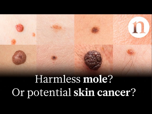
Photos of skin cancer, particularly those captured using dermoscopy, provide magnified views of skin lesions. Healthcare professionals analyze these images to assess characteristics like color, patterns, and borders, aiding in the differentiation between benign and malignant growths.
Question 2: What are the benefits of using photos in skin cancer management?
Photos of skin cancer enable ongoing monitoring of lesion progression and treatment response. They facilitate communication between healthcare providers and patients, supporting informed decision-making and personalized treatment plans.
Question 3: Are photos of skin cancer always accurate?
While photos of skin cancer are valuable diagnostic tools, they may not always provide a definitive diagnosis. Factors like image quality, lesion depth, and individual variations can influence accuracy. A physical examination and biopsy remain essential for confirmation.
Question 4: How can I take effective photos of skin cancer for medical evaluation?
For optimal photos, use natural lighting, focus clearly on the lesion, and capture images from multiple angles. Include a ruler or coin for scale and avoid applying pressure or distorting the lesion.
Question 5: What are the limitations of using photos in skin cancer diagnosis?
Photos of skin cancer may not capture all the necessary information, especially for deep or irregular lesions. They rely on the expertise of the healthcare provider interpreting the images, and may be affected by factors like skin color and hair.
Question 6: How do photos contribute to skin cancer research and prevention?
Photos of skin cancer are invaluable for research, aiding in the study of epidemiology, risk factors, and treatment outcomes. They also support public health campaigns, raising awareness and promoting early detection.
In summary, photos of skin cancer are powerful tools that enhance the diagnosis and management of this prevalent condition. They provide visual documentation, facilitate communication, and support research efforts. However, limitations exist, and a comprehensive approach that includes physical examination, biopsy, and expert interpretation is crucial for accurate diagnosis and effective treatment.
Moving forward, we will delve deeper into the latest advancements in skin cancer imaging and explore how these technologies are revolutionizing skin cancer detection and care.
Tips for Taking Effective Photos of Skin Cancer
To ensure accurate diagnosis and effective management, high-quality photos of skin cancer are essential. Here are some practical tips to help you capture clear and informative images:
Tip 1: Use natural lighting whenever possible, as artificial light can alter the appearance of skin lesions.
Tip 2: Focus clearly on the lesion, avoiding blurry or out-of-focus images.
Tip 3: Capture images from multiple angles to provide a comprehensive view of the lesion.
Tip 4: Include a ruler or coin for scale to help healthcare providers assess the size of the lesion.
Tip 5: Avoid applying pressure or distorting the lesion, as this can affect its appearance.
Tip 6: If possible, take photos before starting any treatment, as this provides a baseline for comparison.
Tip 7: Store and organize photos securely for easy access during follow-up appointments.
By following these tips, you can contribute to accurate diagnosis, effective treatment planning, and improved skin cancer outcomes.
These practical tips empower individuals to take an active role in their skin cancer care, complementing the expertise of healthcare professionals. As we explore the latest advancements in skin cancer imaging in the next section, we will discover how these technologies are further enhancing the accuracy and accessibility of skin cancer detection.
Conclusion
In summary, "photos of skin cancer" have emerged as indispensable tools in the diagnosis, monitoring, and management of this prevalent condition. Through dermoscopy, telemedicine, and artificial intelligence, these photos empower healthcare professionals with detailed visualizations, remote consultations, and automated lesion analysis, leading to improved patient outcomes.
The integration of "photos of skin cancer" into research, public health, and legal documentation further underscores their versatility. They contribute to our understanding of skin cancer epidemiology, support screening programs and awareness campaigns, and provide objective evidence for medical-legal purposes. By capturing the visual characteristics of skin lesions, these photos facilitate accurate diagnosis, informed decision-making, and effective patient care.