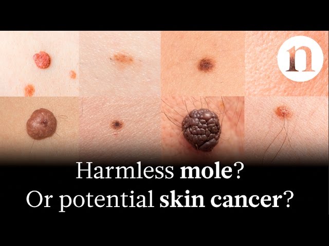
Skin cancer images are visual representations of skin lesions or abnormalities that may indicate the presence of skin cancer. They can be captured through various imaging techniques, such as dermoscopy or photography, and are essential for early detection and diagnosis of skin cancer.
Skin cancer images play a crucial role in healthcare by providing valuable information for medical professionals. They help identify suspicious lesions, track their progression over time, and guide treatment decisions. Historically, the development of non-invasive imaging techniques like dermoscopy has greatly enhanced the accuracy and accessibility of skin cancer detection.
This article delves into the significance of skin cancer images, exploring their applications, benefits, limitations, and ethical considerations in the field of dermatology.
Skin Cancer Images
Skin cancer images are essential for early detection, diagnosis, and treatment of skin cancer. They provide valuable information for medical professionals, helping them identify suspicious lesions, track their progression, and guide treatment decisions.
- Diagnostic
- Non-invasive
- Visual
- Objective
- Representable
- Archivable
- Comparative
- Teledermatologic
- Educational
These aspects highlight the importance of skin cancer images in dermatology. They enable accurate and timely diagnosis, facilitate monitoring of lesions over time, and support informed treatment planning. Additionally, skin cancer images play a crucial role in telemedicine, education, and research, contributing to improved patient care and advancements in the field.
Diagnostic
In the context of skin cancer images, the diagnostic aspect holds paramount importance. It involves the analysis and interpretation of these images to identify suspicious lesions, differentiate between benign and malignant growths, and guide appropriate medical interventions.
- Visual Assessment: Dermatologists and other healthcare professionals visually examine skin cancer images to detect abnormal patterns, colors, shapes, and structures that may indicate the presence of skin cancer.
- Dermoscopic Analysis: Dermoscopy, a non-invasive imaging technique, provides magnified views of skin lesions, allowing for a more detailed examination of their architectural features, patterns, and blood vessels.
- Computer-Aided Diagnosis (CAD): CAD systems utilize machine learning algorithms to analyze skin cancer images, providing objective and quantitative assessments to assist healthcare professionals in making diagnostic decisions.
- Teledermatology: Skin cancer images enable teledermatology consultations, where patients can send images of their skin lesions to dermatologists for remote evaluation, increasing access to specialized care.
These diagnostic facets collectively contribute to the accurate and timely detection of skin cancer, facilitating appropriate treatment and potentially improving patient outcomes. Skin cancer images serve as valuable diagnostic tools, empowering healthcare professionals with the ability to make informed decisions and provide optimal care to patients.
Non-invasive
In the realm of skin cancer detection and diagnosis, the term "non-invasive" holds significant importance in the context of skin cancer images. Non-invasive techniques allow healthcare professionals to examine and analyze skin lesions without causing any harm or discomfort to the patient.
Skin cancer images play a crucial role in non-invasive skin cancer detection. Dermoscopy, a widely used non-invasive imaging technique, utilizes a specialized magnifying lens to capture close-up images of skin lesions. This enables dermatologists to visualize the intricate structures, patterns, and blood vessels within the lesion, aiding in the identification of suspicious features that may indicate skin cancer.
Non-invasive skin cancer images have revolutionized the field of dermatology. They provide a safe and effective means of examining skin lesions, allowing for early detection, accurate diagnosis, and appropriate treatment planning. The non-invasive nature of these techniques has made skin cancer screening more accessible and convenient, leading to improved patient outcomes.
In summary, non-invasive skin cancer images are a cornerstone of modern dermatology. They allow healthcare professionals to examine skin lesions without causing harm to the patient, facilitating early detection, accurate diagnosis, and timely treatment of skin cancer.
Visual
In the context of skin cancer images, the term "visual" holds profound significance. It encompasses the process of examining and interpreting skin cancer images, utilizing the sense of sight to detect, analyze, and characterize suspicious lesions on the skin.
Visual examination is a critical component of skin cancer image analysis. Dermatologists and other healthcare professionals rely on visual cues, such as color, shape, size, and patterns, to identify potential skin cancers. Dermoscopy, a non-invasive imaging technique, enhances visual examination by providing magnified views of skin lesions, allowing for a more detailed assessment of their architectural features and vascular patterns.
Real-life examples of visual examination in skin cancer images include the identification of irregular borders, asymmetry, color variations, and the presence of atypical blood vessels within a lesion. These visual clues can indicate the presence of skin cancer and guide further diagnostic procedures, such as biopsies.
The practical applications of visual examination in skin cancer images are immense. Early detection of skin cancer is crucial for successful treatment and improved patient outcomes. Visual examination of skin cancer images enables the timely identification of suspicious lesions, facilitating prompt referral to specialists and appropriate interventions.
In summary, the visual aspect of skin cancer images is paramount for accurate diagnosis and timely management of skin cancer. Visual examination allows healthcare professionals to detect, analyze, and characterize suspicious lesions, guiding appropriate medical interventions and improving patient outcomes.
Objective
In the context of skin cancer images, "objective" refers to the scientific, unbiased, and reproducible nature of these images, enabling consistent and accurate interpretation by healthcare professionals.
- Standardized Acquisition: Skin cancer images are captured using standardized protocols, ensuring consistency in lighting, magnification, and image quality, allowing for reliable comparisons and analysis over time.
- Digital Format: Digital skin cancer images facilitate easy storage, sharing, and archiving, enabling multiple experts to review and collaborate on the interpretation, leading to more accurate diagnoses.
- Quantitative Analysis: Advanced image analysis techniques, such as computer-aided diagnosis (CAD) systems, provide objective measurements and quantifiable data from skin cancer images, aiding in the assessment of lesion characteristics and risk stratification.
- Interobserver Agreement: The high level of interobserver agreement among trained dermatologists in interpreting skin cancer images indicates the reliability and reproducibility of these images, ensuring consistency in diagnosis and reducing diagnostic variability.
The objective nature of skin cancer images is crucial for ensuring accurate and reproducible diagnoses. Standardization, digital format, quantitative analysis, and interobserver agreement collectively contribute to the clinical utility and reliability of skin cancer images in the detection and management of skin cancer.
Representable
In the context of skin cancer images, "representable" highlights the ability of these images to accurately portray and capture the characteristics of skin lesions, providing valuable information for healthcare professionals to make informed clinical decisions.
- Visual Representation: Skin cancer images provide a visual representation of skin lesions, allowing healthcare professionals to examine the size, shape, color, and other visual features that can indicate the presence of skin cancer.
- Lesion Characteristics: These images can capture important lesion characteristics, such as asymmetry, border irregularity, color variation, and the presence of ulceration or bleeding, which are crucial for accurate diagnosis.
- Disease Progression: Skin cancer images can be used to track the progression of skin lesions over time, allowing healthcare professionals to monitor changes and assess the effectiveness of treatment interventions.
- Educational Value: Skin cancer images serve as valuable educational tools for healthcare professionals and patients alike, illustrating different types of skin cancer and their corresponding clinical features.
The representability of skin cancer images is crucial for early detection, accurate diagnosis, and appropriate treatment planning. These images provide a comprehensive visual representation of skin lesions, enabling healthcare professionals to make informed clinical decisions and improve patient outcomes.
Archivable
Within the realm of skin cancer images, "Archivable" holds immense significance as it pertains to the long-term storage and preservation of these images for future reference and analysis. This aspect ensures the accessibility and integrity of skin cancer images, enabling various benefits and applications.
- Digital Storage: Skin cancer images are predominantly stored digitally in specialized databases or cloud-based systems, ensuring their preservation and easy retrieval for future reference.
- Historical Record: Archived skin cancer images serve as a valuable historical record, allowing healthcare professionals to track the progression of skin lesions over time and monitor the effectiveness of treatment interventions.
- Research and Education: Archived skin cancer images provide a rich resource for research and education, enabling the study of disease patterns, development of diagnostic algorithms, and training of healthcare professionals.
- Legal and Medicolegal: Skin cancer images can serve as legal and medicolegal documentation, providing objective evidence of skin lesions and their changes over time, which is crucial in medico-legal cases or insurance claims.
The archival nature of skin cancer images is essential for comprehensive patient care, research advancements, and medicolegal purposes. It ensures the long-term preservation and accessibility of these images, facilitating informed decision-making, improved patient outcomes, and contributions to the field of dermatology.
Comparative
In the context of skin cancer images, "Comparative" holds significant relevance, as it involves the process of comparing and contrasting skin cancer images to identify similarities, differences, and changes over time. This comparative analysis plays a critical role in the detection, diagnosis, and management of skin cancer.
Comparative analysis of skin cancer images allows healthcare professionals to assess the progression of skin lesions, monitor treatment response, and differentiate between benign and malignant lesions. By comparing current skin cancer images with previous images or with images of known skin cancer types, dermatologists can make informed decisions about the appropriate course of action.
Real-life examples of comparative analysis in skin cancer images include tracking the changes in size, shape, color, and texture of a skin lesion over time to monitor its progression. Additionally, comparing skin cancer images with a database of known skin cancer images can assist in the differential diagnosis of skin lesions, helping to distinguish between benign and malignant growths.
The practical applications of comparative analysis in skin cancer images are vast. It enhances the accuracy of skin cancer diagnosis, optimizes treatment plans, and improves patient outcomes. Comparative analysis also facilitates the study of skin cancer patterns, risk factors, and treatment modalities, contributing to the advancement of dermatological research and practice.
Teledermatologic
In the realm of skin cancer images, teledermatologic aspects play a significant role in the remote evaluation, diagnosis, and management of skin lesions. This involves the transmission of skin cancer images from patients to healthcare professionals using telecommunication technologies.
- Remote Consultation: Teledermatologic services enable patients to consult with dermatologists remotely, allowing for the assessment of skin cancer images without the need for in-person visits.
- Image-Based Diagnosis: Through teledermatology, healthcare professionals can analyze skin cancer images to make diagnostic decisions, providing preliminary assessments and triage recommendations.
- Follow-Up Monitoring: Teledermatologic platforms facilitate the ongoing monitoring of skin lesions over time, allowing healthcare professionals to track changes and provide timely interventions.
- Improved Accessibility: Teledermatology expands access to specialized dermatologic care, particularly in underserved or remote areas where in-person consultations may be limited.
The teledermatologic aspect of skin cancer images revolutionizes patient care by enabling remote evaluation, facilitating early detection, and improving accessibility to expert dermatologic services. It represents a valuable tool for both patients and healthcare professionals, optimizing the management of skin cancer and contributing to improved patient outcomes.
Educational
Educational aspects of skin cancer images hold immense significance in the realm of dermatology and patient care. These images serve as valuable tools for teaching and learning about skin cancer, enabling healthcare professionals, students, and the general public to enhance their knowledge and understanding of this prevalent condition.
"Educational" is a critical component of "skin cancer images" as it facilitates the dissemination and comprehension of essential information related to skin cancer. Through various platforms such as textbooks, medical journals, conferences, and online resources, skin cancer images are widely used to illustrate clinical presentations, histopathological findings, and treatment modalities, aiding in the education and training of healthcare professionals.
Real-life examples of the educational value of skin cancer images can be observed in the training of dermatology residents and medical students. These images provide an invaluable resource for studying different types of skin cancer, their characteristic features, and appropriate management strategies.
The practical applications of educational skin cancer images extend beyond the medical field. Public health campaigns and educational materials utilize these images to raise awareness about skin cancer, promote early detection, and encourage preventive measures such as sun protection. By leveraging the visual impact of skin cancer images, educational initiatives can effectively communicate important messages and empower individuals to take proactive steps in safeguarding their skin health.
Frequently Asked Questions about Skin Cancer Images
This FAQ section addresses common inquiries and clarifies important aspects of skin cancer images.
Question 1: What are skin cancer images?
Answer: Skin cancer images are visual representations of skin lesions or abnormalities that may indicate the presence of skin cancer. They play a crucial role in early detection, diagnosis, and management of skin cancer.
Question 6: How are skin cancer images used in education and research?
Answer: Skin cancer images are valuable educational tools for healthcare professionals, students, and the general public. They are used in textbooks, medical journals, and online resources to illustrate clinical presentations, histopathological findings, and treatment modalities. These images also contribute to research, aiding in the study of skin cancer patterns, risk factors, and treatment outcomes.
In summary, skin cancer images are essential for early detection, diagnosis, monitoring, and education in the field of dermatology. Their applications extend beyond clinical practice, contributing to research and public health initiatives aimed at reducing the burden of skin cancer worldwide.
This comprehensive overview of skin cancer images provides a solid foundation for further discussions on their significance in clinical decision-making, patient care, and ongoing advancements in dermatologic research.
TIPS
This section provides practical tips to optimize the use of skin cancer images in clinical practice, ensuring accurate diagnosis, effective management, and improved patient outcomes.
Tip 1: Utilize standardized image acquisition protocols to ensure consistent lighting, magnification, and image quality, facilitating reliable comparisons and analysis.
Tip 2: Employ advanced image analysis techniques such as dermoscopy and computer-aided diagnosis (CAD) to enhance visualization and objectively assess lesion characteristics.
Tip 3: Implement teledermatology platforms to expand access to expert dermatologic care, particularly in underserved or remote areas.
Tip 4: Integrate skin cancer images into educational materials for healthcare professionals, students, and the general public to promote early detection and preventive measures.
Tip 5: Establish a standardized reporting system for skin cancer images to ensure clear and concise communication among healthcare providers.
Tip 6: Regularly review and update image acquisition and analysis techniques to incorporate advancements in technology and improve diagnostic accuracy.
Tip 7: Foster collaboration between dermatologists and other healthcare professionals to ensure appropriate interpretation and utilization of skin cancer images in multidisciplinary settings.
Tip 8: Engage in research initiatives to contribute to the development of novel image analysis algorithms and improve the understanding of skin cancer patterns and outcomes.
By implementing these tips, healthcare professionals can leverage skin cancer images to enhance patient care, optimize treatment strategies, and contribute to advancements in the field of dermatology.
These practical recommendations serve as a foundation for the concluding section, which will delve into the broader implications and future directions of skin cancer image analysis in clinical practice and research.
Conclusion
In conclusion, skin cancer images have revolutionized the field of dermatology, providing valuable insights into the early detection, diagnosis, monitoring, and management of skin cancer. The use of advanced imaging techniques, telemedicine, and educational resources has significantly improved patient care and outcomes.
Key takeaways include the importance of standardized image acquisition, advanced image analysis, and teledermatology in enhancing diagnostic accuracy and accessibility. Additionally, the integration of skin cancer images into educational materials and research initiatives has contributed to a better understanding of skin cancer patterns and advancements in treatment strategies.