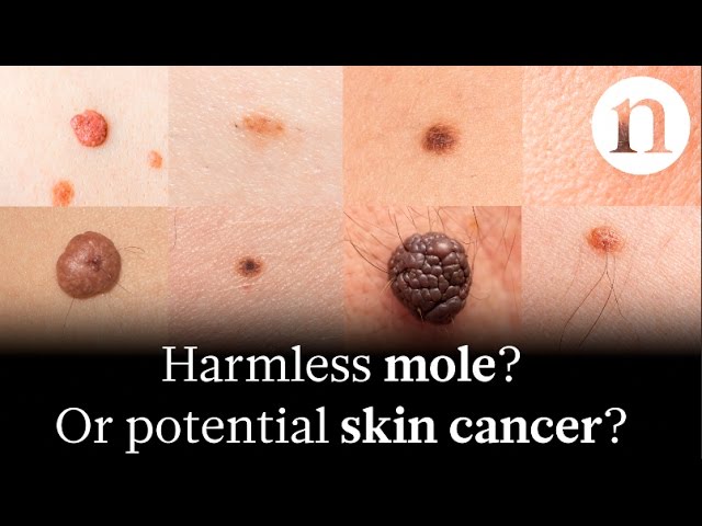
Pictures of skin cancer are visual representations of cancerous lesions on the skin, often used for educational, diagnostic, and research purposes.
These images play a critical role in increasing awareness about skin cancer, aiding in early detection and timely treatment. A significant historical development in this field was the invention of dermoscopy, a non-invasive technique used to magnify and illuminate skin lesions, allowing for better visualization and analysis of suspicious areas.
This article will explore the various types of pictures of skin cancer, their applications, and how they have revolutionized our understanding and approach to this prevalent disease.
Pictures of Skin Cancer
Pictures of skin cancer serve as invaluable tools in multiple aspects, including:
- Diagnosis
- Education
- Research
- Awareness
- Documentation
- Monitoring
- Telemedicine
- Prevention
These images provide irrefutable evidence of cancerous lesions, permitting accurate diagnosis and timely intervention. By illustrating the diverse presentations of skin cancer, pictures educate both healthcare providers and the general public, enhancing awareness and promoting early detection. Moreover, they are essential for research, aiding in the study of disease patterns, treatment outcomes, and the development of novel therapies. Pictures of skin cancer serve as a valuable tool for monitoring disease progression, documenting treatment responses, and facilitating telemedicine consultations, extending access to expert care. Additionally, they play a critical role in prevention, highlighting the importance of regular skin examinations and sun-protective measures.
Diagnosis
Diagnosis is the process of identifying a disease or condition. In the context of skin cancer, pictures play a pivotal role in diagnosis. Visual examination of the skin is a crucial step in the diagnostic process, and pictures allow for a more detailed and objective assessment of suspicious lesions.
Pictures of skin cancer are essential for accurate diagnosis because they provide a permanent record of the lesion's appearance, which can be helpful for tracking changes over time or comparing the lesion to other images. This is particularly important for lesions that are difficult to diagnose based on clinical examination alone. Pictures can also be used to document the response to treatment, which can be helpful for monitoring the effectiveness of therapy and making treatment decisions.
In addition to aiding in the diagnosis of individual patients, pictures of skin cancer are also valuable for research purposes. By studying pictures of skin cancer, researchers can learn more about the different types of skin cancer, how they develop, and how they can be treated. This information can be used to develop new and more effective treatments for skin cancer.
Education
Pictures of skin cancer are not only essential for diagnosis and research, but also for education. They play a crucial role in educating healthcare providers, patients, and the general public about skin cancer, its various forms, and the importance of early detection and prevention.
-
Patient Education
Pictures of skin cancer can help patients understand their condition, treatment options, and prognosis. They can also provide patients with a visual reference for monitoring changes in their skin lesions.
-
Public Awareness
Pictures of skin cancer can be used to raise awareness about the disease and its risk factors. They can be displayed in public places, such as schools, libraries, and community centers, to educate people about the importance of sun protection and regular skin exams.
-
Healthcare Provider Education
Pictures of skin cancer can be used to train healthcare providers to recognize and diagnose skin cancer. They can also be used to illustrate different treatment options and their outcomes.
-
Research Education
Pictures of skin cancer can be used to study the disease and its progression. They can also be used to develop new treatments and prevention strategies.
Overall, pictures of skin cancer are a valuable educational tool that can be used to improve the understanding of skin cancer among patients, the public, and healthcare providers. This can lead to earlier detection, better treatment outcomes, and ultimately, a reduction in the incidence of skin cancer.
Research
In the context of skin cancer, pictures play a pivotal role in research. They provide a wealth of data that can be used to study the disease, its progression, and its treatment. This research has led to significant advances in our understanding of skin cancer, and it continues to play a vital role in the development of new and more effective treatments.
-
Diagnosis
Pictures of skin cancer can be used to develop and validate diagnostic algorithms. These algorithms can help doctors to diagnose skin cancer more accurately, which can lead to earlier treatment and better outcomes.
-
Treatment
Pictures of skin cancer can be used to study the effectiveness of different treatments. This information can help doctors to choose the best treatment for each patient, and it can also help to develop new and more effective treatments.
-
Prevention
Pictures of skin cancer can be used to study the risk factors for skin cancer. This information can help people to take steps to reduce their risk of developing skin cancer.
-
Education
Pictures of skin cancer can be used to educate patients, the public, and healthcare providers about skin cancer. This education can help to increase awareness of skin cancer, and it can also help people to recognize the signs and symptoms of skin cancer.
Overall, pictures of skin cancer are a valuable research tool that has led to significant advances in our understanding of the disease. This research continues to play a vital role in the development of new and more effective treatments for skin cancer.
Awareness
Awareness plays a crucial role in the fight against skin cancer. Pictures of skin cancer can be a powerful tool for raising awareness about the disease, its risk factors, and the importance of early detection. By providing a visual representation of the disease, pictures can help people to understand what skin cancer looks like and how to spot it. This can lead to earlier diagnosis and treatment, which can improve the chances of a cure.
There are many different ways to use pictures of skin cancer to raise awareness. They can be used in educational campaigns, on websites, and in social media. They can also be used in doctor's offices and clinics to help patients understand their condition. In addition, pictures of skin cancer can be used to train healthcare providers to recognize and diagnose the disease.
One example of a successful awareness campaign that used pictures of skin cancer is the "Ugly Duckling" campaign. This campaign used images of skin cancer lesions to educate people about the different types of skin cancer and the importance of early detection. The campaign was successful in raising awareness about skin cancer and led to an increase in the number of people who were screened for the disease.
Pictures of skin cancer can be a powerful tool for raising awareness about the disease and its risk factors. By providing a visual representation of the disease, pictures can help people to understand what skin cancer looks like and how to spot it. This can lead to earlier diagnosis and treatment, which can improve the chances of a cure.
Documentation
Documentation is an integral aspect of pictures of skin cancer. It serves as a permanent record that can be used for a variety of purposes, including patient care, research, and education.
-
Clinical Documentation
Clinical documentation includes the patient's medical history, physical examination findings, and treatment plan. It is essential for providing continuity of care and tracking the patient's progress over time.
-
Research Documentation
Research documentation includes data collected from clinical trials and other research studies. It is used to advance our understanding of skin cancer and develop new treatments.
-
Educational Documentation
Educational documentation includes patient education materials, textbooks, and journal articles. It is used to educate patients, the public, and healthcare professionals about skin cancer.
-
Legal Documentation
Legal documentation includes medical records and other documents that may be used in legal proceedings. It is important for protecting the rights of patients and healthcare providers.
Documentation is essential for the management of skin cancer. It provides a permanent record of the patient's condition and treatment, which can be used for a variety of purposes. It is also important for research and education, and can help to protect the rights of patients and healthcare providers.
Monitoring
Monitoring is the process of observing and tracking a patient's condition over time. It is an essential part of managing skin cancer, as it allows doctors to assess the effectiveness of treatment and detect any changes in the cancer.
Pictures of skin cancer play a critical role in monitoring. They provide a permanent record of the cancer's appearance, which can be used to compare changes over time. This is especially important for cancers that are difficult to diagnose based on clinical examination alone. Pictures of skin cancer can also be used to monitor the response to treatment, which can help doctors to make informed decisions about the best course of action.
One real-life example of monitoring pictures of skin cancer is the use of dermoscopy. Dermoscopy is a non-invasive technique that uses a special magnifying lens to visualize the skin at a higher magnification. This allows doctors to see more detail in the skin, which can help them to identify and diagnose skin cancers at an early stage. Pictures of skin cancer taken with a dermoscope can be used to monitor the cancer over time and assess the response to treatment.
Monitoring pictures of skin cancer is an essential part of managing the disease. It allows doctors to assess the effectiveness of treatment and detect any changes in the cancer. Pictures of skin cancer can be used to monitor the cancer over time and assess the response to treatment.
Telemedicine
Within the realm of skin cancer diagnosis and management, telemedicine, the practice of providing healthcare remotely using telecommunication technology, has revolutionized the utilization of pictures of skin cancer.
-
Remote Consultations
Patients can transmit pictures of suspicious skin lesions to healthcare providers for evaluation and preliminary diagnosis, facilitating timely access to specialist opinions, especially in underserved areas.
-
Dermatoscopic Imaging
Specialized mobile devices or attachments allow for high-quality dermoscopic images to be captured and transmitted, enabling remote providers to assess skin lesions with greater accuracy.
-
Automated Analysis
Artificial intelligence algorithms can analyze pictures of skin cancer, aiding in the identification and classification of suspicious lesions, providing an additional layer of support for remote consultations.
-
Patient Monitoring
Patients can regularly send pictures of their skin lesions for ongoing monitoring, allowing healthcare providers to track changes and intervene promptly if necessary.
These telemedicine applications, by leveraging pictures of skin cancer, expand access to expert dermatologic care, expedite diagnoses, and enhance the overall management of skin cancer.
Prevention
Prevention plays a crucial role in the fight against skin cancer. Pictures of skin cancer can be a powerful tool for raising awareness about the disease, its risk factors, and the importance of prevention. By providing a visual representation of the disease, pictures can help people to understand what skin cancer looks like and how to spot it. This can lead to earlier diagnosis and treatment, which can improve the chances of a cure.
There are many different ways to use pictures of skin cancer for prevention. They can be used in educational campaigns, on websites, and in social media. They can also be used in doctor's offices and clinics to help patients understand their condition. In addition, pictures of skin cancer can be used to train healthcare providers to recognize and diagnose the disease.
One example of a successful prevention campaign that used pictures of skin cancer is the "Ugly Duckling" campaign. This campaign used images of skin cancer lesions to educate people about the different types of skin cancer and the importance of early detection. The campaign was successful in raising awareness about skin cancer and led to an increase in the number of people who were screened for the disease.
Pictures of skin cancer can be a powerful tool for prevention. By providing a visual representation of the disease, pictures can help people to understand what skin cancer looks like and how to spot it. This can lead to earlier diagnosis and treatment, which can improve the chances of a cure.
Frequently Asked Questions about Pictures of Skin Cancer
This section addresses frequently asked questions and clarifies common misconceptions regarding pictures of skin cancer, providing valuable information for better understanding and appropriate action.
Question 1: What types of pictures are used for skin cancer diagnosis?
Answer: Dermatologists utilize various imaging techniques to capture pictures of skin cancer, including clinical photography, dermoscopy, and reflectance confocal microscopy. These specialized methods enhance the visualization and analysis of skin lesions for accurate diagnosis.
Question 2: How do pictures aid in skin cancer treatment?
Answer: Pictures serve as a valuable tool throughout the treatment journey. They facilitate treatment planning by providing detailed documentation of the lesion's characteristics and extent. Additionally, pictures are used to monitor treatment response, assess healing progress, and make necessary adjustments to the treatment regimen.
Question 3: Can pictures of skin cancer be used for self-screening?
Answer: While self-screening using pictures can complement regular skin self-examinations, it should not replace professional evaluation by a dermatologist. Dermatologists possess the expertise and experience to accurately interpret skin cancer pictures and provide appropriate guidance.
Question 4: How can I obtain high-quality pictures of my skin lesions?
Answer: For optimal picture quality, ensure adequate lighting and use a high-resolution camera. Position the camera perpendicular to the skin lesion and maintain a consistent distance. Avoid using flash, as it can create glare and distort the lesion's appearance.
Question 5: What are the limitations of using pictures for skin cancer diagnosis?
Answer: While pictures provide valuable information, they have limitations. Subcutaneous features of lesions, such as depth and vascularity, may not be fully captured in pictures. Additionally, image quality and interpretation can vary depending on the photographer's skills and experience.
Question 6: How can I securely share pictures of my skin lesions with healthcare providers?
Answer: Utilize secure online platforms or patient portals designated for sharing medical information. These platforms encrypt and protect patient data, ensuring confidentiality and privacy.
These FAQs provide a concise overview of essential information regarding pictures of skin cancer. Understanding these aspects empowers individuals to actively participate in their skin health management and seek appropriate medical attention when needed.
In the following section, we will delve deeper into the role of artificial intelligence in skin cancer detection, exploring its capabilities and potential impact on skin cancer diagnosis and management.
Tips for Taking Pictures of Skin Cancer
To obtain high-quality pictures for accurate diagnosis and monitoring, follow these practical tips:
Tip 1: Choose the Right Lighting
Ensure adequate and natural lighting to avoid shadows and distortions.
Tip 2: Position the Camera Properly
Hold the camera perpendicular to the skin lesion, maintaining a consistent distance.
Tip 3: Avoid Flash
Flash can create glare, obscuring the lesion's true appearance.
Tip 4: Use a High-Resolution Camera
Higher resolution captures more detail, aiding in accurate assessment.
Tip 5: Focus on the Lesion
Ensure the camera focuses on the skin lesion, not the surrounding area.
Tip 6: Include a Ruler or Coin
Place a ruler or coin next to the lesion to provide scale for size evaluation.
Tip 7: Take Multiple Pictures
Capture images from different angles and distances to provide a comprehensive view.
By following these tips, you can obtain high-quality pictures that facilitate accurate diagnosis and effective management of skin cancer.
These pictures, along with other diagnostic tools and a dermatologist's expertise, contribute to early detection, appropriate treatment, and improved outcomes for skin cancer patients.
Conclusion
In summary, "pictures of skin cancer" play a pivotal role in the diagnosis, monitoring, research, education, and awareness of skin cancer. They provide a valuable tool for healthcare providers and individuals alike, contributing to early detection, appropriate treatment, and improved patient outcomes. Key points include the use of pictures in clinical documentation, telemedicine, and prevention campaigns, showcasing their versatility and impact on skin cancer management.
As we advance, the integration of artificial intelligence and machine learning in skin cancer detection holds promising potential to further enhance accuracy and accessibility. The future outlook for pictures of skin cancer is bright, with continued advancements in imaging technology and AI algorithms revolutionizing skin cancer diagnosis and management.