Primary malignant melanoma of the oral cavity is rare d. Oral malignant melanoma is commonly diagnosed in men aged 51 60 years whereas it is commonly diagnosed in females aged 61 70 years.
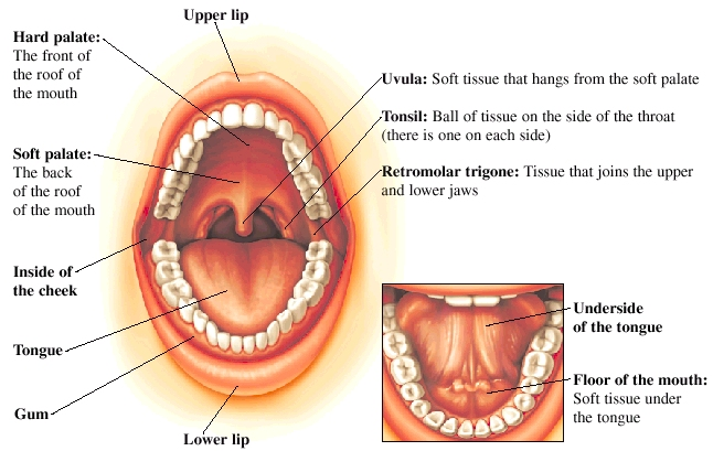
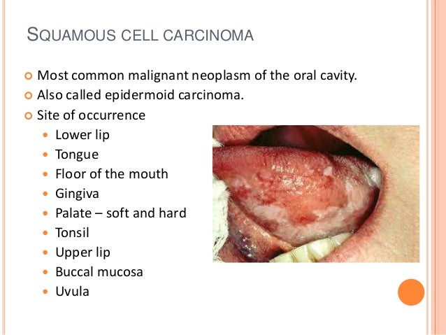
malignant oral cancerous lesions are most frequently located on the is important information accompanied by photo and HD pictures sourced from all websites in the world. Download this image for free in High-Definition resolution the choice "download button" below. If you do not find the exact resolution you are looking for, then go for a native or higher resolution.
Don't forget to bookmark malignant oral cancerous lesions are most frequently located on the using Ctrl + D (PC) or Command + D (macos). If you are using mobile phone, you could also use menu drawer from browser. Whether it's Windows, Mac, iOs or Android, you will be able to download the images using download button.
It has a benign counterpart known as the benign melanoma b.

Malignant oral cancerous lesions are most frequently located on the. What are the 5 types of. What is the most common type of oral cancer. Most malignant melanomas arise on the skin as a result of prolonged exposure to chemicals such as benzene c.
What do we need to do with premalignant or potentially malignant lesions. Opscc are the palatine tonsils and the lingual tonsils located on the base of tongue. Most cancerous lesions are derived from potentially malignant oral disorders pmod.
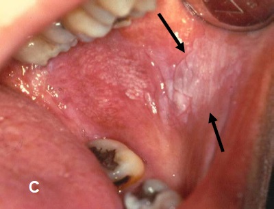
Diagnosis of oral ulcerative lesions might be quite challenging. Leukoplakia erythroplakia actinic cheilitis submucous fibrosis and lichen planus. Erythroplakia is defined as any lesion of the oral.
The most common intraoral location is the tongue. Potentially malignant lesions of the oral cavity are most likely to undergo malignant transformation to ocscc in the following sites. The world health organization who points out the following lesions as the main pmod.
The average patient age at diagnosis is 56 years. Which of the following statements about malignant melanoma is true. In the mouth it most commonly starts as a painless white patch that thickens develops red patches an ulcer and continues to growwhen on the lips it commonly looks like a persistent crusting ulcer that does not heal and slowly grows.
Tongue lateral borders ventral tongue base dorsum. This narrative review article aims to introduce an updated decision tree for diagnosing oral ulcerative lesions on the basis of their diagnostic features. The evidence that oral leukoplakias are pre malignant is mainly derived from follow up studies showing that between 1 and 18 of oral pre malignant lesions will develop into oral cancer.
Oral malignant melanoma is largely a disease of those older than 40 years and it is rare in patients younger than 20 years. Squamous cell carcinoma 90 of oral cancers. Although erythroplakia is an infrequent oral condition its risk of malignant progression is the highest among all oral precancerous lesions.
What is the mean time for scc to develop from. Oral cancer also known as mouth cancer is cancer of the lining of the lips mouth or upper throat. Various general search engines and specialized databases including pubmed.
Oral cancer and potentially cancerous lesions early detection and diagnosis 97 and the sensitivity and specificity values from existing literature a ppv of 127 and a false. Leukoplakias are white plaques or spots that cannot be removed by scraping and these lesions arent characterized clinically or. Most common oral lesions.
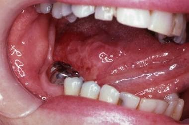 Cancers Of The Oral Mucosa Background Pathophysiology
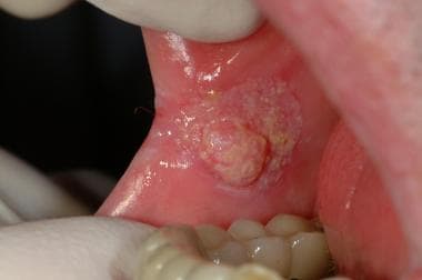 Cancers Of The Oral Mucosa Clinical Presentation History
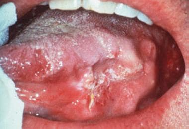 Cancers Of The Oral Mucosa Background Pathophysiology
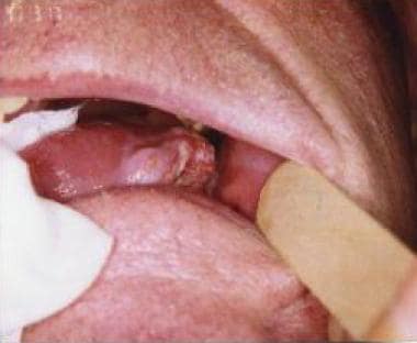 Malignant Tumors Of The Mobile Tongue Practice Essentials
 Common Oral Lesions Part Ii Masses And Neoplasia
Oral Mucosal Ulceration A Clinician S Guide To Diagnosis
 Common Oral Lesions Part Ii Masses And Neoplasia
